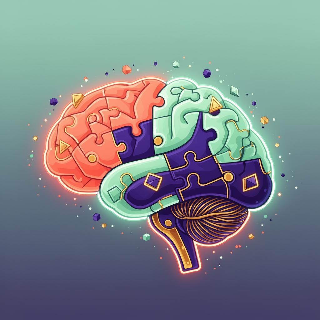
The cerebral cortex - the wrinkled outer layer - is divided into four lobes: frontal (planning, decision-making, voluntary movement), parietal (touch, spatial awareness, navigation), temporal (hearing, language, long-term memory), and occipital (vision). Beneath sits the cerebellum, which fine-tunes movement and contributes to attention and learning. The Society for Neuroscience's BrainFacts.org and the NIH's National Institute of Neurological Disorders and Stroke (NINDS) both publish freely available primers maintained by working researchers.
The prefrontal cortex - the very front of the frontal lobe - is the seat of executive function: working memory, inhibition, and planning. It is the last region to fully mature, typically not finishing until the mid-20s (Sowell et al., Nature Neuroscience, 2003, UCLA), which is why adolescents and young adults can be brilliant and impulsive in the same minute.
The hippocampus, tucked inside each temporal lobe, is essential for forming new episodic memories. The famous London taxi driver studies by Dr. Eleanor Maguire at University College London (PNAS, 2000) showed that the posterior hippocampus is measurably larger in drivers who have memorized the city's 25,000 streets - direct evidence that adult brains physically remodel themselves through use.
The amygdala drives rapid threat detection and emotional salience; the insula represents internal body states; the basal ganglia coordinate habit and reward. Together these regions form the limbic and reward circuits that shape motivation. Damage or dysregulation in any node - well documented by the NIH BRAIN Initiative and Allen Institute for Brain Science - produces distinct, recognizable clinical patterns.